
Skeleton Pose Stock Photo Download Image Now iStock
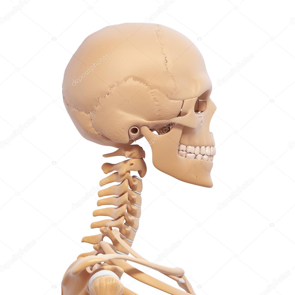
The skull is the skeletal structure of the head that supports the face and protects the brain. It is subdivided into the facial bones and the cranium, or cranial vault ( Figure 7.3.1 ). The facial bones underlie the facial structures, form the nasal cavity, enclose the eyeballs, and support the teeth of the upper and lower jaws.
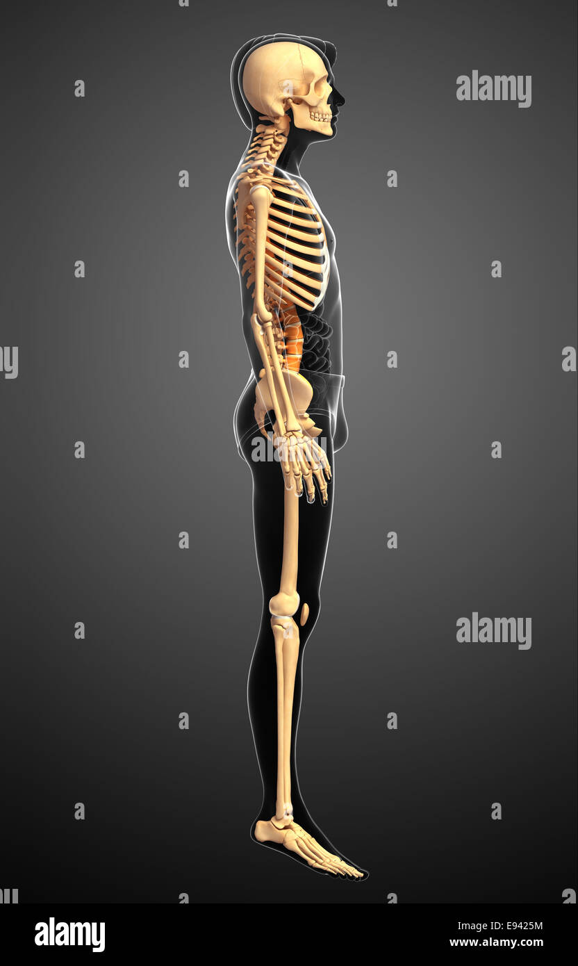
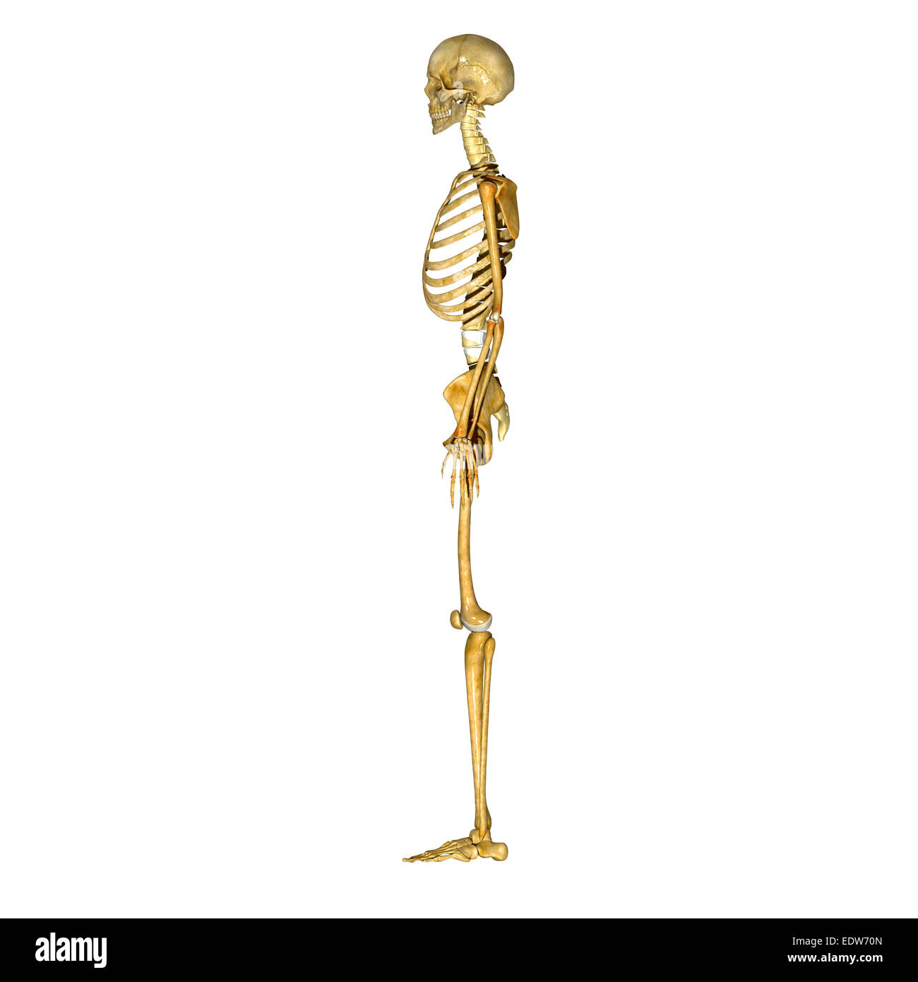
side view standing skeleton of human body Stock Photo 66107008 Alamy
Bone is living tissue that makes up the body's skeleton. There are 3 types of bone tissue, including the following: Compact tissue. The harder, outer tissue of bones. Cancellous tissue. The sponge-like tissue inside bones. Subchondral tissue. The smooth tissue at the ends of bones, which is covered with another type of tissue called cartilage.
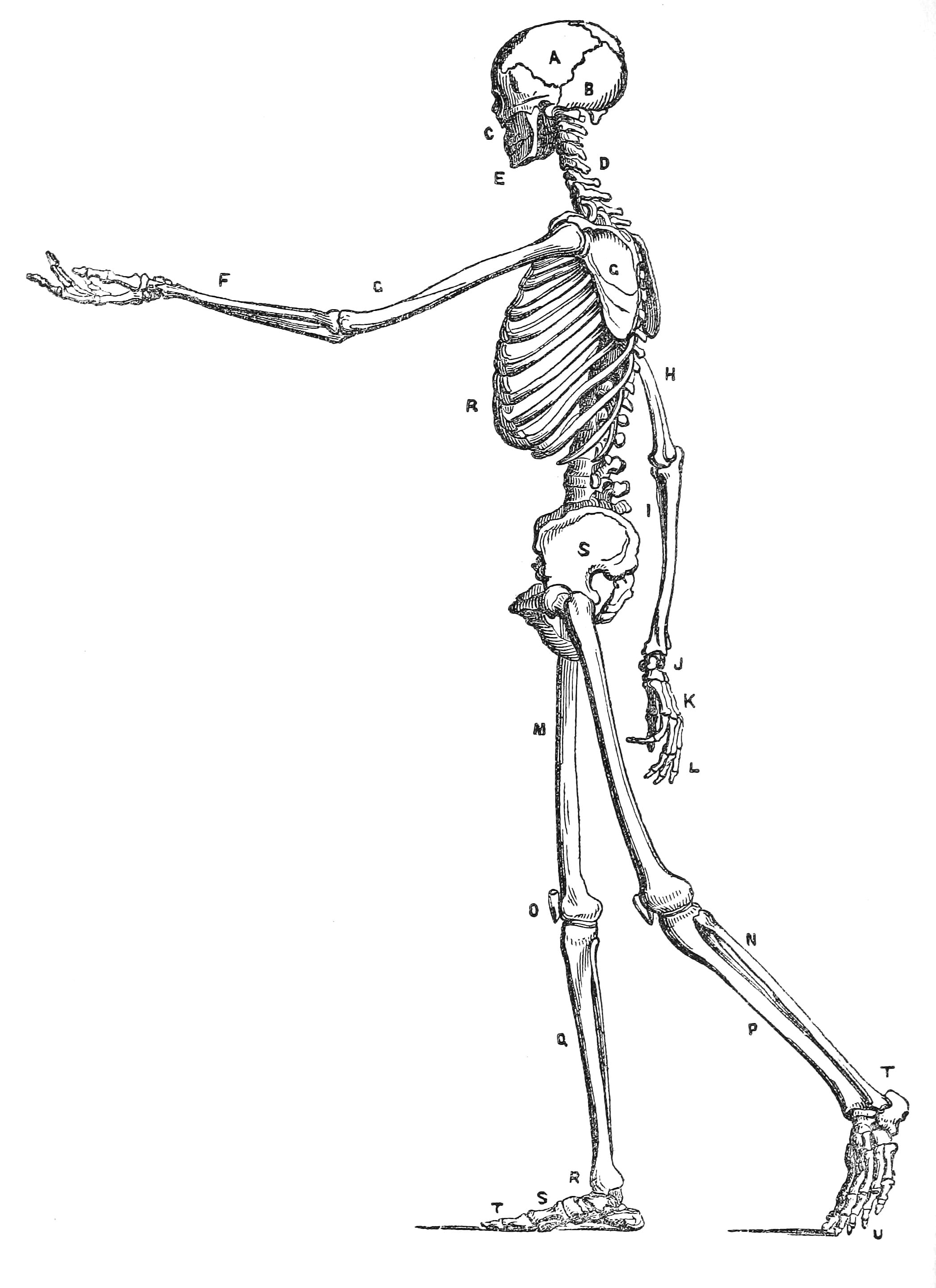
Human Skeleton Drawing Anatomy Art Surface View
The greater wing of the sphenoid is a structure that extends from the side of the body of the bone to curve backwards and laterally. The most posterior part of this projection is a triangular process that fits neatly into the ridge between the petrous and squamous portion of the temporal bone. The sphenoid bone (lateral view)
Human skeleton side view — Stock Photo © pixologic 22677793
Dec. 24, 2023, 4:25 AM ET (Yahoo News) Human skeletons, remains of sharks, blood-sucking bats. human skeleton, the internal skeleton that serves as a framework for the body. This framework consists of many individual bones and cartilages.

skeleton Skeleton, Stock images, Dark side
Spine Structure and Function. Your spine is an important bone structure that supports your body and helps you walk, twist and move. Your spine is made up of vertebrae (bones), disks, joints, soft tissues, nerves and your spinal cord. Exercises can strengthen the core muscles that support your spine and prevent back injuries and pain.
Skeleton side view hires stock photography and images Alamy
The Anatomy of the Skeletal System By Aubrey Bailey, PT, DPT, CHT Published on May 23, 2023 Medically reviewed by Geetika Gupta, MD The skeletal system comprises 206 bones and has two main parts—the axial skeleton and the appendicular skeleton.

Human skeleton front and side view men anatomy Vector Image
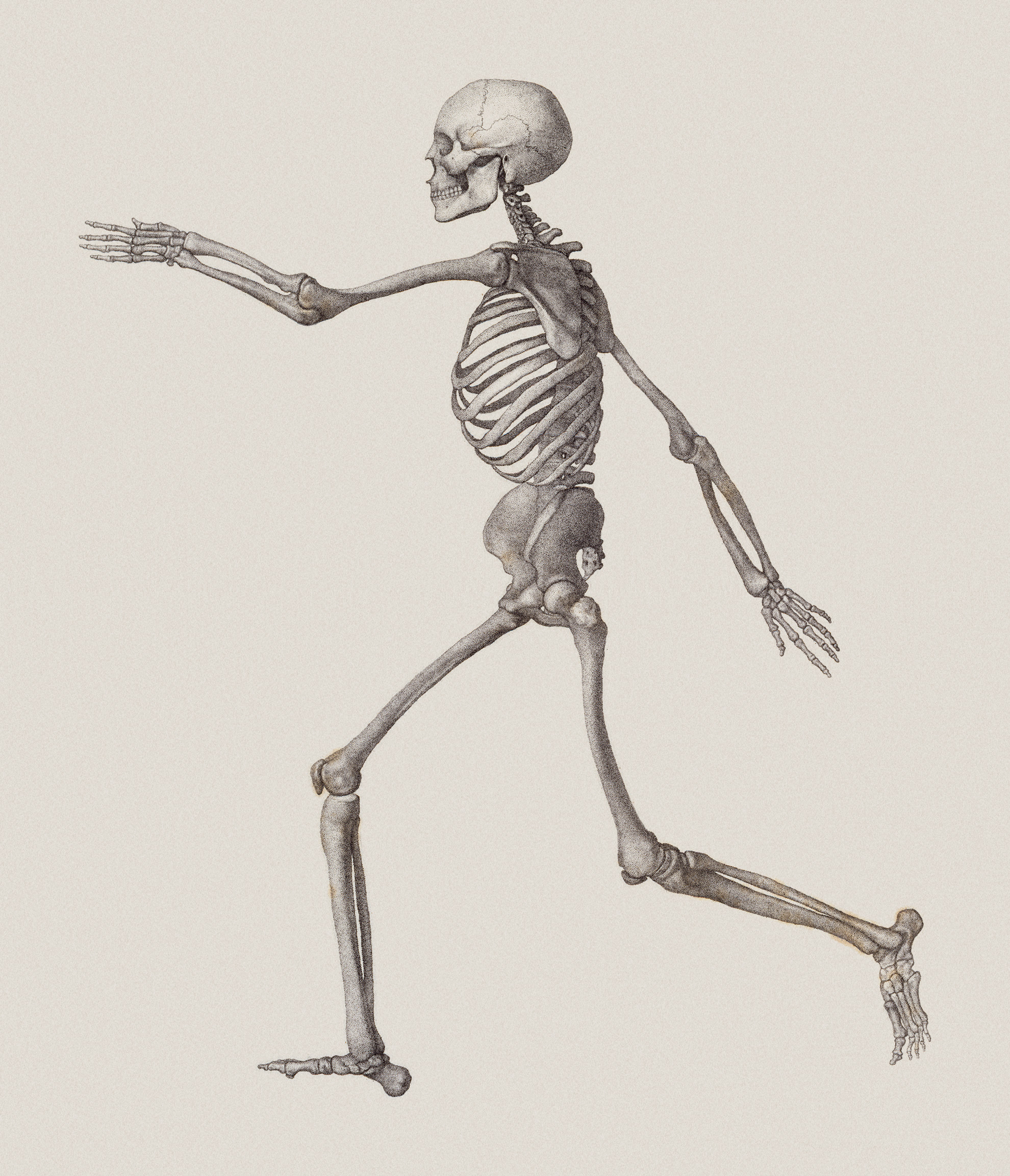
What does the skeletal system do? At the simplest level, the skeleton is the framework that provides structure to the rest of the body and facilitates movement. The skeletal system includes over 200 bones, cartilage, and ligaments. Read on to get 10 key facts about the human skeleton. 1. The Skeletal System Consists Of More Than Bones

Skeleton anatomy pose sketch Skeleton drawings, Skeleton art
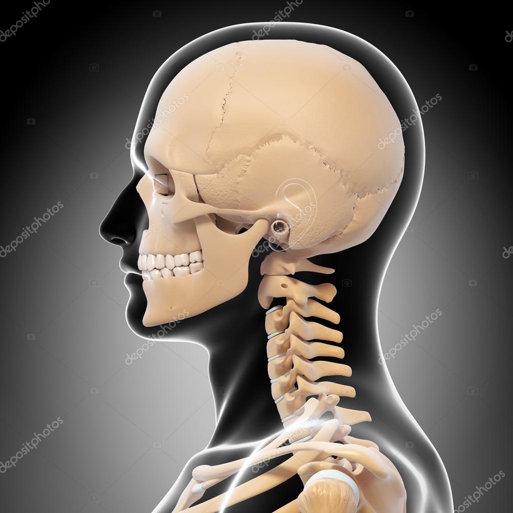
The cranium (skull) is the skeletal structure of the head that supports the face and protects the brain. It is subdivided into the facial bones and the brain case, or cranial vault ( Figure 7.3 ). The facial bones underlie the facial structures, form the nasal cavity, enclose the eyeballs, and support the teeth of the upper and lower jaws.
Front Side View Skeleton High Resolution Stock Photography and Images
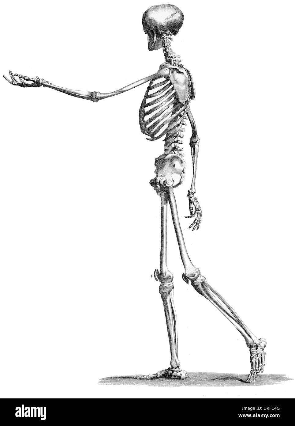
about the human skeleton and identify some of the important features of our skeletal anatomy. This document contains an outline of an adult human standing 187 ½ cm tall (or 6'2").. side of the body. 4. Compare the height of the skeleton on the printout with the printout of Lucy and your own height.

Brandon Jackson's Portfolio Studying the Human Skeleton
The skeleton is made up of 206 bones in the adult and contributes to the form and shape of the body. The skeleton has several important functions for the body. The bones of the skeleton provide support for the soft tissues. For example, the rib cage supports the thoracic wall.

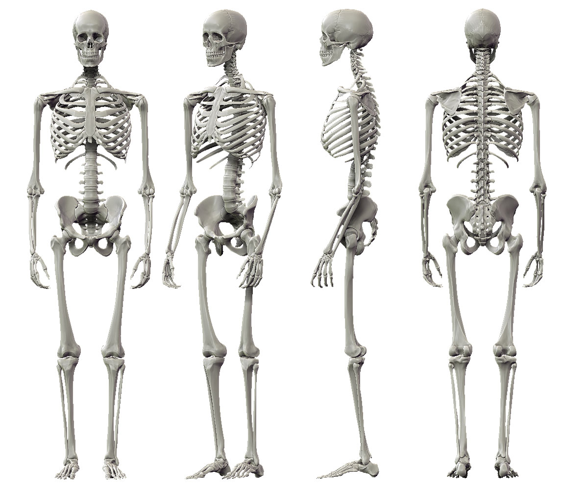
Illustration of anterior and posterior views of human skeletal
Skull, skeletal framework of the head of vertebrates, composed of bones or cartilage, which form a unit that protects the brain and some sense organs. The skull includes the upper jaw and the cranium.. The atlas turns on the next-lower vertebra, the axis, to allow for side-to-side motion. inferior view of the human skull. internal surface of.
Vintage Human Anatomy Illustration Skeleton Side View Free Vintage
Browse 4,100+ human skeleton side view stock photos and images available, or search for human anatomy to find more great stock photos and pictures. human anatomy Sort by: Most popular The human body and a skeleton with a silhouette of a body. A male, A vector illustration on a white background. Human male skeleton full figure. Five angle views.
Molly's Art Blog A Bit of Character Work...
There are three auditory ossicles on each side of the head, known as the: malleus (hammer) incus (anvil) stapes (stirrup) They work together to transmit sound waves from the surrounding.
Human skeleton side view Stock Photo by ©pixologic 22677875
When to see a doctor Summary The foot is an intricate part of the body, consisting of 26 bones, 33 joints, 107 ligaments, and 19 muscles. Scientists group the bones of the foot into the phalanges,.

Skeleton Side View stock vector. Illustration of bones 11827135
The human skull consists of 22 bones (or 29, including the inner ear bones and hyoid bone) which are mostly connected together by ossified joints, so called sutures.The skull is divided into the braincase (neuro cranium) and the facial skeleton (viscerocranium).Its main task is the protection of the most important organ in the human body: the brain.

Human skeleton side view Vector Image 1585226 StockUnlimited
Endoskeleton. An endoskeleton is a skeleton that consists of hard, mineralized structures located within the soft tissue of organisms. An example of a primitive endoskeletal structure is the spicules of sponges. The bones of vertebrates are composed of tissues, whereas sponges have no true tissues (Figure 38.1. 1 ).
